Immunity needs a platform for enriching communication.
The apparent randomness in our immune systems is mainly a result of genetic and environmental complexity. Beside this intrinsic randomness, however, the changes in the morphology of the immune cell and the localization of the molecules involved in immunity, when an organism senses a pathogen (virus, bacteria, …), usually cause well-defined patterns. These patterns, known as synapses, are dynamic, polarized macromolecular structures formed at the interface of two cells when they come into close opposition to communicate. The communication is through signal transition and is specially important when our body is threatened by a foreign substance which provoke an immune response. The foreign substances (pathogens) drive some protein fragments (peptide) the recognition of which is critical for the initiation of the immune response.
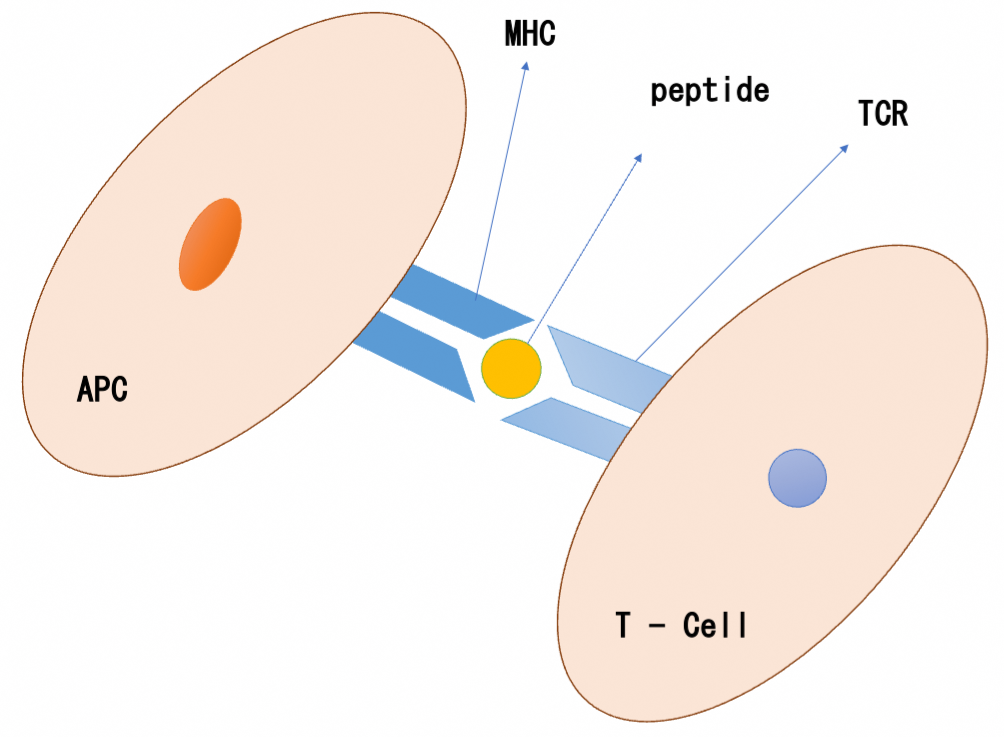
T-cell, an essential part of the adaptive immune system, is responsible for the recognition of these peptides. Another cell named as antigen presenting cell (APC) bind these peptides and present them to T-cell [1] (schematically shown in the figure).
The presenting and recognition of pathogen’s fragments (peptides) respectively by APC and T-Cell (as shown in the figure above) is just the initial step of what we call immune response. Some molecules on the surface of two cells, T-Cell receptor (TCR) molecule and its binding partner on the opposite cell (APC) named as peptide major histocompatibility complex (pMHC), are responsible for the initial signaling and the communication at the interface. In addition, Other molecules called adhesion molecules facilitate, as their name suggests, the activation of T-Cell as well as the contact of T-Cell and APC. The spatial and temporal movement of these surface molecules generally results in the formation of the immune synapse (IS) in the area between two cells; A platform for enriching signaling molecules and controlling T- Cell activation.
Two forms of Immune Synapses (IS)
After formation of micro-clusters by bound TCR and pMHC molecules, these clusters fuse to the center of the synapse and form the central supramolecular activation cluster (cSMAC) in the center of the synapse. Meanwhile, the adhesion molecules interact with each other, surrounding the cSMAC, and form the peripheral supramolecular activation cluster (pSMAC). The resulted structure is a form of a bullseye for the immune synapse.
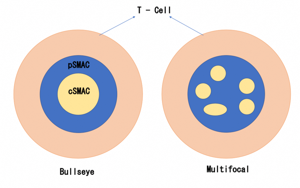
Another possible form of the synapse is a multifocal IS and it is observed when the micro-clusters of TCR-pMHC scatter at the multiple sites at the interface instead of fusing to the center. Both bullseye and multifocal structures of IS are depicted in the figure.
In the process of IS formation, it is not just the spatial movement of the molecules that matters, but also their temporal movement should correlate with the T-Cell activation. In other words, the motility of the molecules from the surface, where they are uniformly distributed, to a place, where they can perform their assigned task, is well orchestrated. That is to say, being at the right place in the right time is necessary for the molecules to act efficiently.
Does different forms of IS have anything to do with its function?
As mentioned earlier, Immune Synapse is believed to be a platform for enriching signaling molecules and controlling T-Cell activation, and consequently immune response. Therefore, different forms of IS must act differently regarding the signaling transmission. In fact, studies showed a higher level of calcium response in multifocal IS than in bullseye IS. Another important function of the IS is killing the infected or tumor cells, and bullseye IS is the main synapse type for this function. In general, these forms can be alternated to each other. A changed structure from multifocal to the bullseye IS is a reflection of a phase transition between initiation and quenching of the T-Cell activation.
Why is it important to study the IS structures?
It is so far clear that the formation of Immune Synapses and the T-Cells activation are critical steps in immunity. Studies showed that, with a high probability, the type and specificity of antigen determines which type of IS is formed. In other words, by knowing the target, T-Cell and surface molecules decide in which form they accumulate. From this point of view, the type of synapse structure is a critical factor in immunotherapy, especially in confronting the antigens from a virus that recognize the adhesion molecules and penetrate into the cell to disrupt the movement of molecules and the formation of the Synapse. In such cases, understanding the structure and function of IS and the mechanism by which IS is formed plays a key role in immunotherapy [2].
References:
[1] “TCR Recognition of Peptide-MHC-I: Rule Makers and Breakers”, Christopher Szeto, et al.
[2] “Immunological Synapse Molecules”, Wei Lin and Zhichao Fan.
